Humans are multicellular organisms.
Multicellular organisms have evolved significantly based on how efficiently they interact with their surrounding environment and communicate with other cells. It is said that the human body contains approximately 37 trillion cells. This is an astonishing number that is hard to comprehend. The fact that so many cells operate in a coordinated and organic manner, much like an orchestra, is truly marvelous and impressive. The very act of detecting change is a sign of life. For lifeless objects, change is meaningless. When various external stimuli are recognized, the body quickly determines how to respond within a very short period of time, and specific responses are executed at the cellular level. The most appropriate and effective response is devised in an incredibly brief moment. For this, close signaling and communication between cells are essential. Without it, maintaining homeostasis, which is necessary for healthy bodily functions, would be challenging. If there is a problem in any part of the signaling system, it can lead to disease, and for that reason, specific processes of the signaling system can also be targeted to treat diseases.
Cells are both transmitters and receivers of signals. Isn't the sum total of accumulated responses that unfold little by little but continuously over the long period of thousands or millions of years evolution? In understanding the fundamental functions of the body's tissues or organs, a clear understanding of these cell-to-cell communication methods and the intracellular signaling system (cell signaling) will certainly be a solid foundation.
Type of signal
A signal can be defined as an external stimulus applied to a cell. It is a signal that causes the cell to perform the necessary actions in response to these stimuli. Let's broadly divide signals into three types.
1. Physical signals: There are physical signals such as light, temperature, pressure, and voltage. For example, skin cells have special receptors that are sensitive to changes in temperature, called thermoreceptors, and can detect and respond to hot or cold stimuli in the surrounding environment. Usually, this information is converted into signals and transmitted to the brain, but there are also receptors that respond directly at the cellular level.
2. Chemical signals: These include chemical molecules such as neurotransmitters, hormones, growth factors, cytokines, etc.
3. Mechanical signal: This is a signal sent by mechanical stimuli such as pressure or shear stress applied to the cell membrane. When stretching, compression, or changes in tension are applied to cells or tissues, the cell membrane can be deformed, potentially compromising the integrity of the cell. Physical contact with neighboring cells can also apply mechanical forces to the cell membrane. Mechanosensitive ion channels present in the cell membrane detect these stimuli.
The receptors are the points at which cells detect the various stimuli described above. Each type of signal is recognized by specialized receptors, and after several steps, the signal ultimately leads to the targeted response.
How do cells react when they receive a signal message ?
When cells detect changes in the external environment through their sensory organs, they will immediately attempt to adjust their internal environment to quickly adapt to these changes for survival. If, for any reason, a large number of cells die or are in a state of stress, the cells will perform cell division to increase their numbers and replenish the dead cells. Hematopoietic stem cells in the bone marrow will differentiate and prioritize the speed at which they produce red blood cells, neutrophils, and platelets based on various signals from the body. Also, if a cell is severely damaged and no longer contributes to survival or is no longer needed in a changed environment, it will send out a signal called apoptosis to remove itself. In other words, when cells receive signals from the outside, they respond to them by responding to them in various ways, such as survival, growth, differentiation, and death. Of course, there are undoubtedly countless reactions that are even more specific, diverse, and complex than can be listed. This is because the response to one stimulus or one signal message is not limited to one, but can be differentiated in countless different ways. Moreover, depending on the location or role of the cell receiving the signal, the same signal can elicit very different responses. As always, the scale of the events occurring at the molecular level within cells seems to be beyond our imagination.
Representative signaling molecules: hormones
One of the representative signaling molecules that the human body produces in response to external stimuli and transmits to cells is hormones. In fact, while studying in detail the major hormones, including thyroid hormones, I realized that to understand how these hormones are directly delivered to cells and the pathways through which they exert their effects, it is essential to first grasp the intracellular signaling mechanisms. Even the same hormone can have different roles in various tissues, so when instructing cells to perform specific tasks, I wanted to understand what pathways the signals take and how they differ. Above, I examined the various types of stimuli that can be recognized from the outside. Signaling pathways involve processes that occur between cells or within the cells themselves.
Signaling pathways
The series of interactions between molecules that occur from the starting point of a signalto its ultimate response in the cell can be collectively referred to as a "pathway." This pathway often includes numerous processes that wake up various enzymes that are inactive and switch them on to an active state. Just like a relay race, where the baton is passed along to reach the finish line, there are chain reactions that follow one after another.
It may seem obvious, but the roles that cells perform are complex and intricate, and so the pathways involved are also highly diverse and detailed. When reading scientific papers related to physiology, one encounters various pathways that sound extremely complex, such as MAPK, ERK, and Ark pathways. However, all these intricate pathways share common processes and procedures. Fortunately, in the field of cell biology, which extensively studies these areas, there seems to be a convention for categorizing and simplifying these pathways to make them more understandable for ordinary people like myself. Nothing in nature is completely disconnected or lacks a cause-and-effect relationship. I intend to explore individual pathways in more detail when the opportunity arises, but for now, I want to understand how cells transmit signals through their common elements.
Distance of signals
Cells respond to a variety of signals. One of the most representative forms is endocrine signaling, where hormones are secreted from glands located somewhere in the body and delivered to target cells or tissues via the bloodstream or lymphatic fluid. Hormones such as thyroid hormones or insulin are often considered long-distance signals because the glands that secrete them and the cells that receive these signals are usually not in close proximity. In contrast, substances secreted by salivary glands, sweat glands, or digestive glands affect only nearby tissues or cells without entering the bloodstream, classifying them as short-distance signals. For reference, hormones are classified as the endocrine system, while sweat glands, salivary glands, etc. fall under the exocrine system.
If a small wound occurs in our body, the injured tissue rapidly sends signals to surrounding cells to secrete histamine. This is a wise immunological response that dilates blood vessels, allowing white blood cells to arrive quickly to prevent infection. Platelets, known for stopping bleeding, release platelet-derived growth factor (PDGF) to promote angiogenesis at the wound site and also send out signals called epidermal growth factor (EGF) to aid in healing and the growth of new skin. These signals are examples of paracrine signaling, which are transmitted locally to surrounding cells or tissues through the interstitial fluid, without needing to enter the bloodstream. The prefix "para" means "near" or "surrounding."
In rare cases, cells may send signals only to themselves (autocrine signaling) or to other cells of the same type. For example, T cells produce autocrine signals when exposed to specific antigens, activating and proliferating themselves to regulate and maintain immune responses. Unfortunately, cancer cells can also exploit this mechanism to proliferate.
Finally, there are instances where signals are transmitted directly between adjacent cells. This can be observed in the heart, where muscle cells send electrical signals to neighboring cells in an orderly fashion, causing them to contract simultaneously.

Signal transmission in neurons
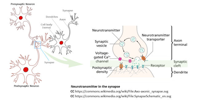
However, when discussing signal transmission, what cannot be left out is the signal between nerve cells, or neurons. The signaling in neurons can be described as a system where electricity and chemistry coexist. The process begins with the action potential, an electrical signal generated by the difference in charge between the inside and outside of the cell membrane. This action potential is rapidly transmitted along the axon, which can be thought of as an electrical wire. When this signal reaches the end of the axon, or the terminal of the neuron, it causes the opening of calcium ion channels in response to the voltage, allowing calcium ions to flow into the neuron. As the concentration of calcium inside the neuron increases, it triggers the release of neurotransmitters stored in vesicles into the extracellular space. At this point, the electrical signal has been converted into a chemical signal.
Why is this necessary? Although neurons may appear to be in contact with each other, there is actually a very small gap between them called the synapse. Since electrical signals cannot be transmitted across this physically non-contacting space, the electrical signal is transformed into a chemical substance, the neurotransmitter, to continue the signaling process. In fact, these synapses play a crucial role in modulating the signals as they travel from the initial source to their final destination. If there is an issue with the signal, it is likely that adjustments will be made at these synapses to address the problem.
Signal Transmission from Pre-Synaptic to Post-Synaptic Neurons
The neurotransmitters secreted by the pre-synaptic neuron, the neuron that sends the signal, into the synaptic space bind to the receptor sites on the cell membrane of the post-synaptic neuron, the neuron that receives the signal on the other side. This binding opens ion channels in the post-synaptic neuron, allowing ions to flow into the cell. Sodium (Na+) and potassium (K+) are cations, while chloride (Cl-) is an anion, and each of these ions has its own specific channels. When the ion channels open, ions rush into the post-synaptic neuron, affecting the charge difference across the cell membrane. Depending on the function of the neurotransmitter, different receptors may be activated, leading to the opening of specific ion channels. This results in the generation of excitatory post-synaptic potentials (EPSPs), which increase the likelihood of triggering an action potential, or inhibitory post-synaptic potentials (IPSPs), which decrease the likelihood of action potential generation.
If channels for cations like Na+ or calcium (Ca²+) open significantly, a large influx of positive ions will cause the membrane potential to shift from negative to positive, reaching the threshold to generate an action potential (excitatory). Conversely, if chloride channels or channels for cations like K+ open due to concentration gradients, positive ions may exit the cell, further increasing the negativity inside the cell and preventing the generation of an action potential (inhibitory). Thus, the type of ion channels that open determines the strength of the electrical potential that can continue the signal to the next neuron. The human body maintains a delicate balance between excitatory and inhibitory potentials to regulate and stabilize the activity of the nervous system. Glutamate is a well-known excitatory neurotransmitter, while gamma-aminobutyric acid (GABA) is a prominent inhibitory neurotransmitter. Other notable neurotransmitters include acetylcholine, norepinephrine, serotonin, and dopamine, all of which play significant roles in the central nervous system.
It is important to recognize that the signaling pathways in the nervous system also involve receptors and channels, which will be explored in greater detail in subsequent discussions.
Articles on ion channels
Articles on action potential generation
The best of the best: Cell membrane
Let’s take a moment to recall the composition of the cell membrane. While studying cells, there are moments that make you want to exclaim, “Wow!”—times when you feel a shiver down your spine because of the sheer complexity and brilliance of biological structures. Personally, learning about the structure of the cell membrane has often left me in awe. Water constitutes about 70% of the human body, but where exactly is all this water? It primarily exists in the intracellular and extracellular fluids.
In this aqueous environment, the cell membrane delineates the boundaries between individual cells, allowing each cell to maintain its own identity. This boundary is formed by the plasma membrane, which is so thin that it can only be seen under an electron microscope. The lipid bilayer that makes up the cell membrane features hydrophilic heads that face the external aqueous environment, while the hydrophobic tails curl inward, creating a dual-layered structure. The outer layer interacts well with the aqueous environment, while the inner layer helps maintain the boundary of the cell.
The head regions on the outside of the membrane interact with both the external environment and the cytoplasm, which is also aqueous. The cell membrane acts as a formidable barrier, filtering what can enter the cell and what cannot, thus providing a robust defense mechanism. It also enables the cell to confine specific molecules to certain compartments. This design is truly remarkable. So, how does a cell, with its well-established boundaries, communicate with the outside world?
While the lipid bilayer forms the fundamental structure and framework of the cell membrane, most of the membrane's functions are carried out by the various proteins embedded within it. Key membrane proteins associated with signal transduction include transport proteins, which encompass various ion channels, membrane receptors, and membrane enzymes. Among these, transport proteins are particularly important, and I will address them in a separate article along with a detailed examination of ion channels.
Now, let’s dive into the signal transduction system. In the following articles, we will explore the intracellular signaling processes and transformations that begin when signaling molecules bind to receptors on the cell membrane's surface. We will start by focusing on the receptors, which serve as the initial contact points for receiving signals.


